The Subdepartment of Histology with the Course of Cytology and Embryology was established in 1932. At that time, the subdepartment had seven microscopes, sixty histological specimens, and two educational tables.
In 1933, the subdepartment received a separate premises (forty square meters). Z.F. Guseva, a collaborator of the subdepartment, played a significant role in organizing and improving teaching. In 1937, Z.F. Guseva was appointed head of the subdepartment. The same year, the first assistant lecturers, A.V. Kirillicheva and I.S. Novic, began working at the subdepartment.
At that time, the subdepartment’s staff also began performing research. Z.F. Guseva researched embryonal pathomorphology under the guidance of Moscow Professor V.K. Beletsky, and A.V. Kirillicheva conducted experimental transplantation of the cornea under the guidance of Professor V.P. Roshin.
During the Great Patriotic War, the staff of the subdepartment expanded due to lecturers who were evacuated from central medical universities. From 1941 to 1945, the subdepartment was headed by L.I. Falin, who arrived from Smolensk. The following lecturers joined the subdepartment: V.V. Anisimova, M.N. Rinchina, and E.D. Piskunova. Under L.I. Falin’s leadership, the educational process was reorganized, and subjects like evolutionary histology and embryology were taught in greater depth.
Professor L.I. Falin delivered excellent lectures and was an exacting examiner. He paid great attention to the improvement of the professional skills of the collaborators. All lecturers in the subdepartment performed research, and in 1943, some defended their theses. L.I. Falin defended his doctoral thesis on the subject “Morphology and Pathogenesis of Teratoid Tumors of the Gonads,” where he proposed a new original theory on the origin of tumors from gonocytes. Lecturer A.V. Kirillicheva defended her Candidate’s dissertation on “Transplantation of the Cornea in Autoplasty Conditions.” In 1944, V.V. Anisimova defended her Candidate’s dissertation on the morphological features of the peripheral nervous system. In 1945, E.D. Piskunova defended her Candidate’s dissertation on “Morphological Changes in Lymph Nodes in Tuberculosis.”
After the end of the Great Patriotic War and the re-evacuation, the subdepartment staff changed, and young former graduates of the Kazakh Medical Institute filled vacant posts, including E.P. Smirnova, N.N. Beliayeva, and T.S. Pliaskina. At that time, the subdepartment was headed by E.G. Troitskaya, who had previously worked in the subdepartment of pathological anatomy. She focused on improving methodological work. The subdepartment also moved to a new educational building, which provided more lecture rooms, and the number of tables, histological specimens, and microscopes increased.
Research work in the subdepartment continued on various topics. E.G. Troitskaya studied the morphological features of primary tuberculosis in adults, while Z.F. Guseva continued her research and, in 1950, defended her Candidate’s dissertation. In 1958, N.N. Beliayeva defended her Candidate’s dissertation on the “Age-related Structural Features of Human Kidneys During Postnatal Life.”
In 1957, Lecturer N.A. Urina joined the subdepartment, having previously worked in the subdepartment of anatomy. In 1960, Lecturer H.H. Musabayeva also joined, having worked in the subdepartment of biology.
From September 1959, after the retirement of Senior Lecturer E.G. Troitskaya, Senior Lecturer A.V. Kirillicheva was elected head of the subdepartment. She led the subdepartment until 1971, with a focus on both methodological and scientific work. The subdepartment’s research topics included “General Regularities of Morphogenesis and Regeneration.”
In 1960, A.V. Kirillicheva defended her doctoral dissertation on “Dynamics of Morphological Changes in Organs and Tissues in Cachexia and Restoration of the Organism, and the Influence of Certain Pharmaceutical Substances on These Processes.” In 1962, Lecturer H.H. Musabayeva defended her Candidate’s dissertation on the influence of ascorbic and nicotinic acids on experimental silicosis. In 1964, Lecturer E.P. Smirnova defended her Candidate’s dissertation on age-related changes in the spleen of laboratory animals.
A.V. Kirillicheva paid great attention to scientific work with students. Three of her postgraduate students defended their Candidate’s dissertations: R.I. Yui (“Cytological and Cytochemical Investigations of the Human Pancreas During Ontogenesis”), M.S. Kalinina (“Influence of Methionine on the Reactive Properties of Connective Tissue During Ontogenesis”), and G.V. Fedorovskih (“Morphological and Histochemical Changes in the Kidneys Due to the Influence of Some Extreme Factors”).
From 1971 to 1983, Senior Lecturer H.H. Musabayeva led the subdepartment. During this time, research focused on the influence of vitamin D deficiency, amino acid imbalance, and balanced nutrition on histogenesis and the structural features of internal organs. Senior Lecturer R.I. Yui conducted research on the combined effects of radiation and dynamic factors on morphological and functional changes in internal organs, which culminated in a doctoral dissertation in 1988.
From 1983 to 1998, Professor A.S. Tolybekov, who had previously defended his Candidate’s and Doctoral dissertations at the Institute of Experimental Medicine in Leningrad, headed the subdepartment. Under his leadership, research began on the influence of environmental factors on the organism, particularly phosphorus pollution. Two doctoral and two Candidate’s dissertations were defended on this subject.
In 1989, the Kazakh National Medical University began teaching in Kazakh. Senior Lecturers R.B. Abyldinov and K.Sh. Baymukhambetov began delivering lectures and lessons in Kazakh. Other lecturers, including K.E. Sisabekov, Zh.O. Ayapova, M.G. Adylova, N.M. Tusupova, M.Zh. Ergazina, Z.N. Dzhangeldina, B.K. Kaniyev, and G.K. Yesimova, also began teaching in Kazakh, and several textbooks on Histology were published in the language.
In 1998, Professor R.I. Yui became head of the subdepartment, where he led research on “Cytological Aspects of Diurnal Rhythms of Internal Organs in Changing Habitats” and “Reactivity of the Oral Mucosa in Normal and Pathological Conditions.” Four Candidate’s dissertations were defended on these topics, and in the 2003-2004 academic year, the subdepartment was recognized for its outstanding methodological work.
In 2013, the university adopted a modular teaching system, and the subdepartment was divided into the Subdepartment of Histology and the Histology-2 module. Senior Lecturer M.Zh. Ergazina was appointed head of the Subdepartment of Histology.
In 2017, the textbook “Fundamentals of Histology of the Oral Cavity and Teeth” was published in English by R.I. Yui, and the “Explanatory Dictionary of the Main Histological Terms in Three Languages” was published by Abildinov R.B., Ergazina M.Zh., and Yui R.I.
Collaborators of the subdepartment have published more than one thousand scientific investigations, including atlases, three textbooks, twenty-six monographs, and manuals. Six doctoral theses and twenty-three Candidate’s dissertations have been defended. The subdepartment regularly participates in international symposiums and conferences.
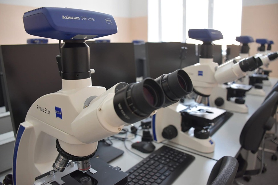
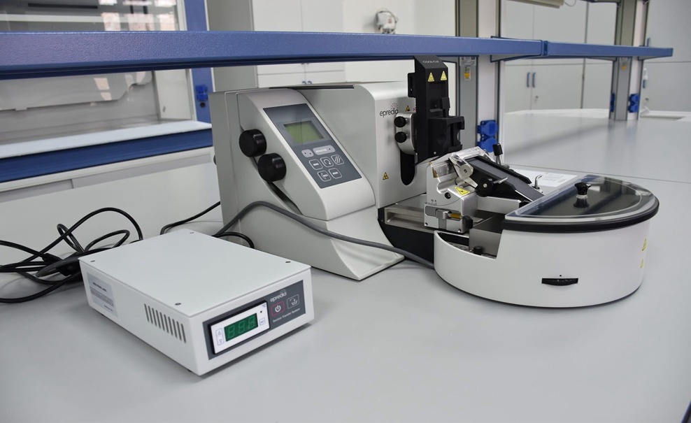
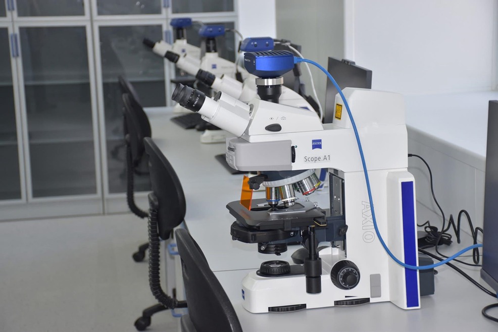
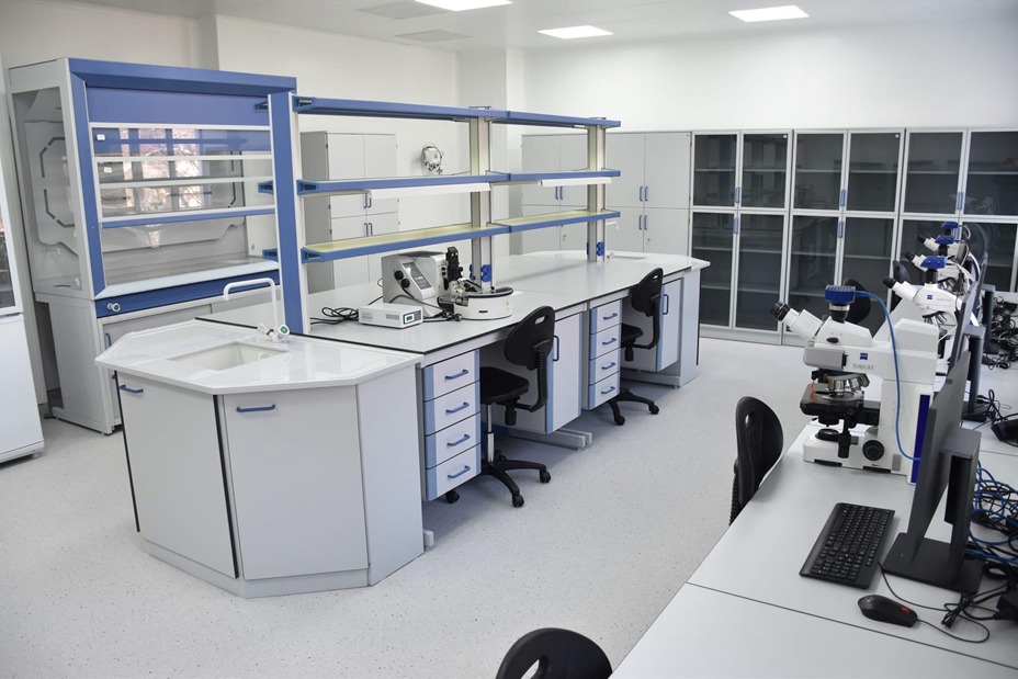
In the 2024-2025 academic year, a grand opening ceremony for the new laboratory of the Department of Histology took place. The department’s laboratory has been transformed into a modern educational space, equipped with all the necessary tools: microscopes, laboratory tables, thermostats, a refrigerator, and many other essential items. This modernization opens up new opportunities for the comprehensive training of students. The future of histological education at KazNMU is closely linked to training provided by Karl Zeiss. Histology department faculty have received specialized training from Karl Zeiss as part of the ‘Modern Histology Laboratory’ project. As a result, the department has successfully integrated new microscopes and equipment into its work. Along with the creation of the histology laboratory, a new-generation digital classroom has been established. The digital classroom is equipped with 12 ‘Primo Star’ microscopes and one ‘Axilob 5’ for the instructor. The advantage of the digital classroom lies in its ability to control and analyze images during interactive lessons through ‘Labscope.’ This setup combines both individual and group-based learning approaches.
New technologies make the study of histology both visual and effective.